
Vitreo-Retinal Surgery
A group of conditions treated with a vitrectomy (an operation to remove the vitreous)
What conditions does this include?
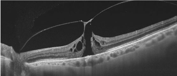
Macula Hole
This occurs when the retina lifts away from the back wall of the eye, and can cause a sudden loss of vision if left untreated. It is treated urgently with surgery to reattach the retina and preserve sight.
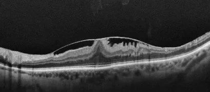
Epiretinal Membrane
An epiretinal membrane is a thin layer of scar tissue that forms on the surface of the retina. As it contracts, it can wrinkle or pucker the retina, causing blurred or distorted vision. Removing the membrane during a vitrectomy allows the retina to settle and vision to improve.
Retinal Detachment
This occurs when the retina lifts away from the back wall of the eye, and can cause a sudden loss of vision if left untreated. It is treated urgently with surgery to reattach the retina and preserve sight.
Intraocular Lens Exchange
Occasionally, a lens implanted during previous cataract surgery needs to be repositioned or exchanged – for example, if it has become dislocated. This can be performed using a vitrectomy approach.
Vitreous floaters
Floaters are small clumps or strands within the vitreous gel that cast shadows on the retina, often seen as spots, threads or cobwebs drifting across your vision. We would normally only consider offering surgery if floaters have been present for at least 6 months and are significantly affecting your quality of life. As with any operation on the eye, patients need to be aware of the small risks involved with surgery.
What to expect on the day of your assessment?
At your assessment, you will have a full examination of your eyes. We will use dilating drops, which will blur your vision for around 3 hours, and we will take a detailed retinal scan (OCT scan).
What does a vitrectomy involve?
The operation is a day procedure, and you normally are in hospital for no more than 2-3 hours in total. The vast majority of retinal surgeries are carried out with the patient awake under local anaesthetic (injection around the eye), which ensures the operation isn’t painful. If you feel you wouldn’t be able to tolerate that, there is an option of having a general anaesthetic.
The operation itself is similar to keyhole surgery where we remove your vitreous and perform any additional procedures necessary.
Are there any risks to the surgery?
Retinal surgery has a high success rate but any operation inside the eye has a degree of risk. These include:
Visual loss due to an infection or severe bleed (1 in 1000 cases)
Risk of retinal detachment
Transient complications such as elevated pressure or inflammation inside the eye which is normally treated with extra drops
Persistent distortion following the surgery